Secreted Peptides: During infection and colonization, pneumococcal cells are organized into complex bacterial communities, termed biofilms. Within these structures, pneumococci produce and receive signals to sense the host environment and communicate across cells. These signals are akin to a bacterial language, and our goal is to decipher their molecular meaning and their role in pathogenesis.
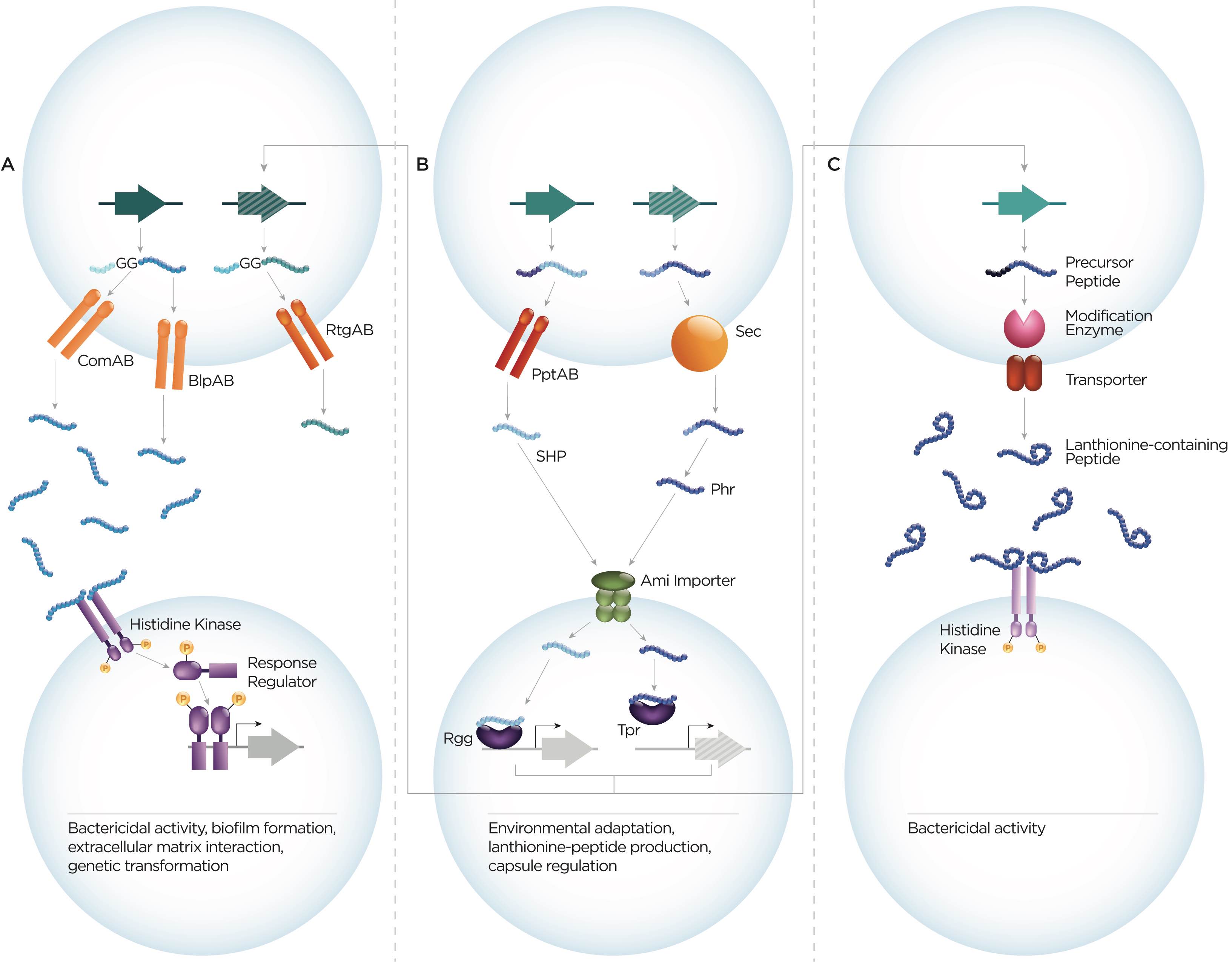
Three main classes of pneumococcal secreted peptides. Schematic showing peptide features, processing and export, receptors and phenotypic consequences of different families of peptides, namely, (A) double-glycine peptides, (B) peptides signaling via the RRNPP superfamily of regulators, and (C) lanthionine-containing peptides. Our lab studies peptides from all three classes, currently we are focused on VP1 and BriC, multiple SHPs and PhrA1 and PhrA2, and LanA. Image by Rachel Kenney. Image from Aggarwal et al, Review in PLoS Pathogens 2020.
Baterial Extracellular Vesicles: In addition to communicating via secreted peptides, bacteria also release information in the form of extracellular vesicles (EVs) that package hundreds of proteins. Until recently, it was assumed that the thick pneumococcal cell wall rendered it incapable of EV release. Our studies join a growing body of work that strongly supports the observation that the pneumococcus releases EVs. We study the role of pneumococcal EVs in gene transfer, inter-strain communication and host interactions.
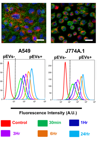
Pneumococcal EVs are internalized by host cells. Top: Representative confocal images of A549 lung epithelial cells (left panels) and murine macrophage (right panels) exposed to bacterial EVs after 24h. Red: pkH26 stained EVs, green: F-actin, blue: DAPI nuclear stain. Botton: Flow cytometry measurements of the amount of DiD label detected in cells before addition of EVs (control) and at five time points post-addition of 20 µg/ml of pneumococcal EVs. The red line demonstrates the fluorescence intensity of cells without any EVs. The vast majority of cells exhibited EV internalization by 24h.